This new bone defect classification was developed because in current classifications the metaphysis and diaphysis are not rated. However, these zones are important for sufficient fixation of the implant in revision total knee arthroplasty (TKA). Therefore, this classification facilitates quantification of bone defects, and supports adequate pre-operative planning of revision TKA. In this manual, the bone defect classification is explained in text and with example video’s.
Description of the classification
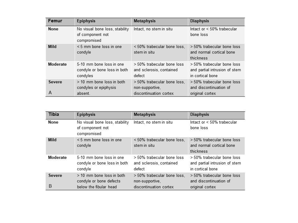
A bone defect is defined as absence of normal bone, including presence of implant, cement, radiolucent lines, and osteolytic lesions. The bone defect classification consist of four rating options for bone defects (none, mild, moderate, severe), that are rated separately per zone (epiphysis, metaphysis, and diaphysis) for both the femur (Figure 1a) and tibia (Figure 1b).

Figure 1a & 1b: Bone defect classification for the femur and for the tibia.
Epiphysis, Metaphysis, Diaphysis
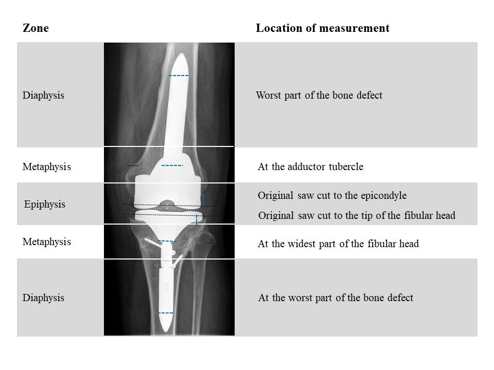
The epiphysis is defined from the original saw cut to the epicondyle (femur) or until the tip of the fibular head (tibia). The bone defect of metaphysis is rated at the adductor tubercle (femur) or at the widest part of the fibular head (tibia). The diaphysis is measured at the worst part of the bone defect, this is usually, but not necessarily, at the tip of the stem. In figure 2 you see an image with the different zones (blue lines) and where in the zone you should measure the bone defect (black dotted line). It is advised to use a ruler in the radiology software to measure the width of the bone defect with regard to the width of the bone to determine whether the bone defect consist of more or less than 50% of the metaphysis or diaphysis. When the AP view and lateral view show a different grade bone defect, the worst grade should be reported.

Figure 2: This shows the definition of femoral and tibial zones used for rating bone defects. The white lines indicate the cutoff points for the zones. The dotted lines (blue) indicate where the measurements of the bone defects for the specific zones should be taken. The anatomic landmarks for the measurements per zone are described on the right and indicated in the picture (black lines).
Instruction video’s
Here you can find a demonstration of the bone defect rating on X-ray for five different patients: